The middle coat of the cornea is composed of collagen fibers and fibroblasts, while the inner surface is simple squamous epithelium.

The outer surface of the cornea is composed of non-keratinized stratified squamous epithelium. The cornea is transparent and covers the colored iris. The fibrous tunic is the outermost layer and consists of the anterior cornea and the posterior sclera. Anatomically, the wall of the eyeball is composed of three layers: the fibrous tunic, the vascular tunic, and the retina. The rest of the eyeball is protected by the orbit into which it fits. The adult eyeball measures about 2.5 centimeters in diameter and only one-sixth of the eyeball is exposed to the outside air. Together, these muscles are capable of moving the eye in almost any direction. The extrinsic eye muscles extend from the bony orbit to the sclera of the eye and are surrounded by a significant amount of periorbital fat.

This is why the nose gets stuffy when an individual cries. The nasolacrimal duct carries lacrimal fluid into the nasal cavity. The two lacrimal canals lead to the lacrimal sac and then into the nasolacrimal duct. From there, the tears enter two small openings called lacrimal punta, and then they pass into two ducts knowns as the lacrimal canals. The lacrimal glands, each about the size of an almond, secrete the lacrimal fluid that drains into six to 12 excretory ducts that empty the fluid onto the surface of the conjunctiva of the upper eyelid. The lacrimal apparatus produces and drains lacrimal fluid, or tears, from the eye. It does not cover the cornea, which is a transparent region that forms the outer anterior surface of the eye. The conjunctiva is a thin, protective mucous that covers the sclera of the eye. The tarsal plate is composed of connective tissue that gives support to the eyelids. The whitish material that sometimes accumulates in that region is from these glands. The lacrimal caruncle is the small, pink, globular spot at the inner corner, or the medial canthus, of the eye. The space between the upper and lower eyelid that exposes the eyeball is the palpebral fissure. Twitches of the eyelids are a common and usually harmless symptom that can be caused by stress and fatigue. Beneath this layer is a donut-shaped muscle that encircles the eye called orbicularis oculi, which is innervated by the 7th cranial nerve, and is in charge of closing the eyelids. The upper eyelid exhibits a greater range of movement than the lower eyelid as it contains the levator palpebrae superioris muscle, a muscle innervated by cranial nerve 3 in charge of lifting the eyelid. The eyelids, also known as the palpebrae, cover the eyes during sleep, protect the eyes from excess light and possibly objects, and spread lubricating secretions over the eyeball. įunctions of the Accessory Structures of the Eye

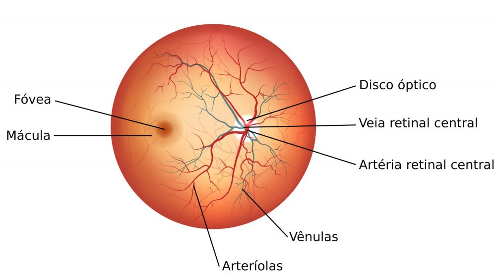
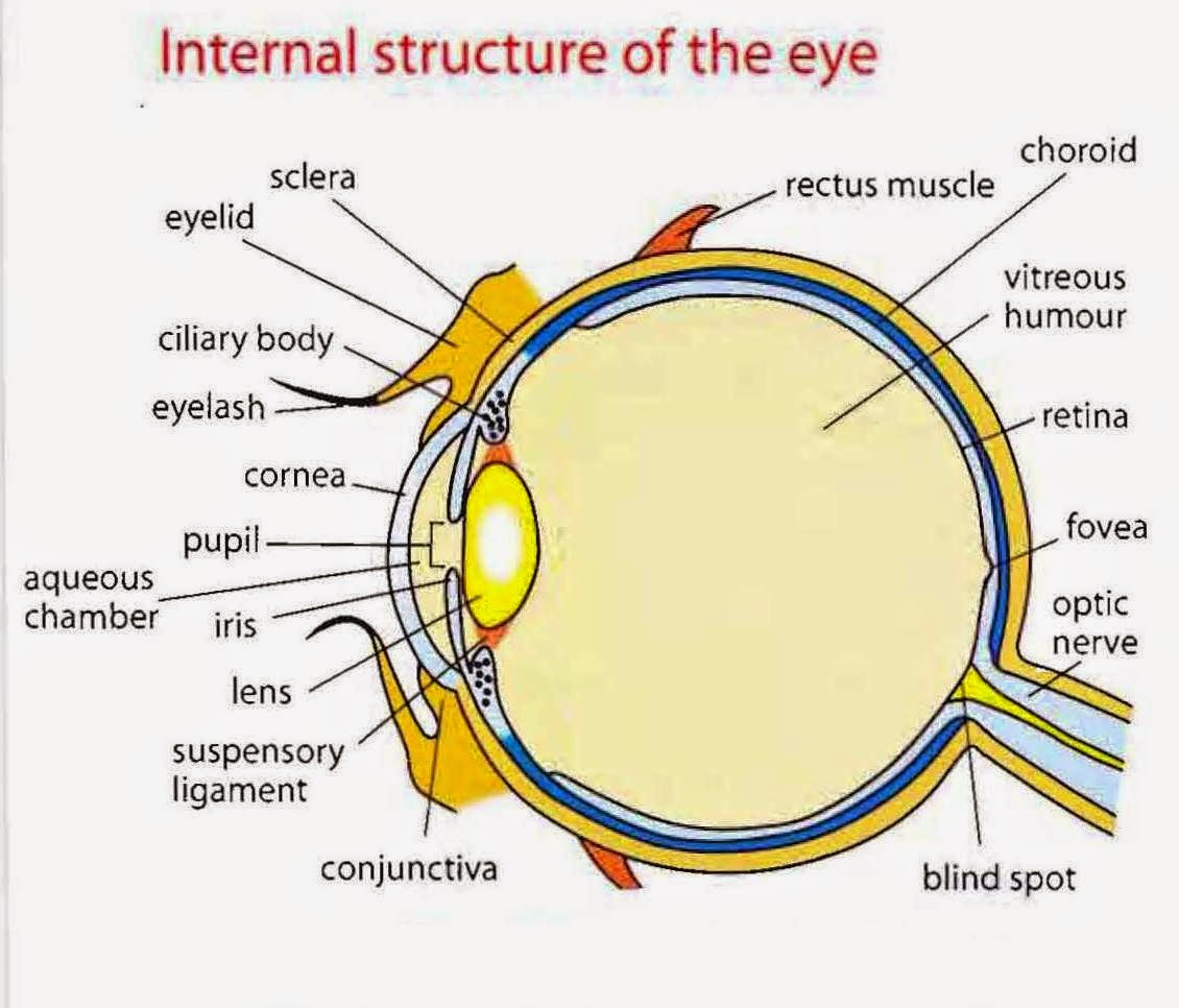
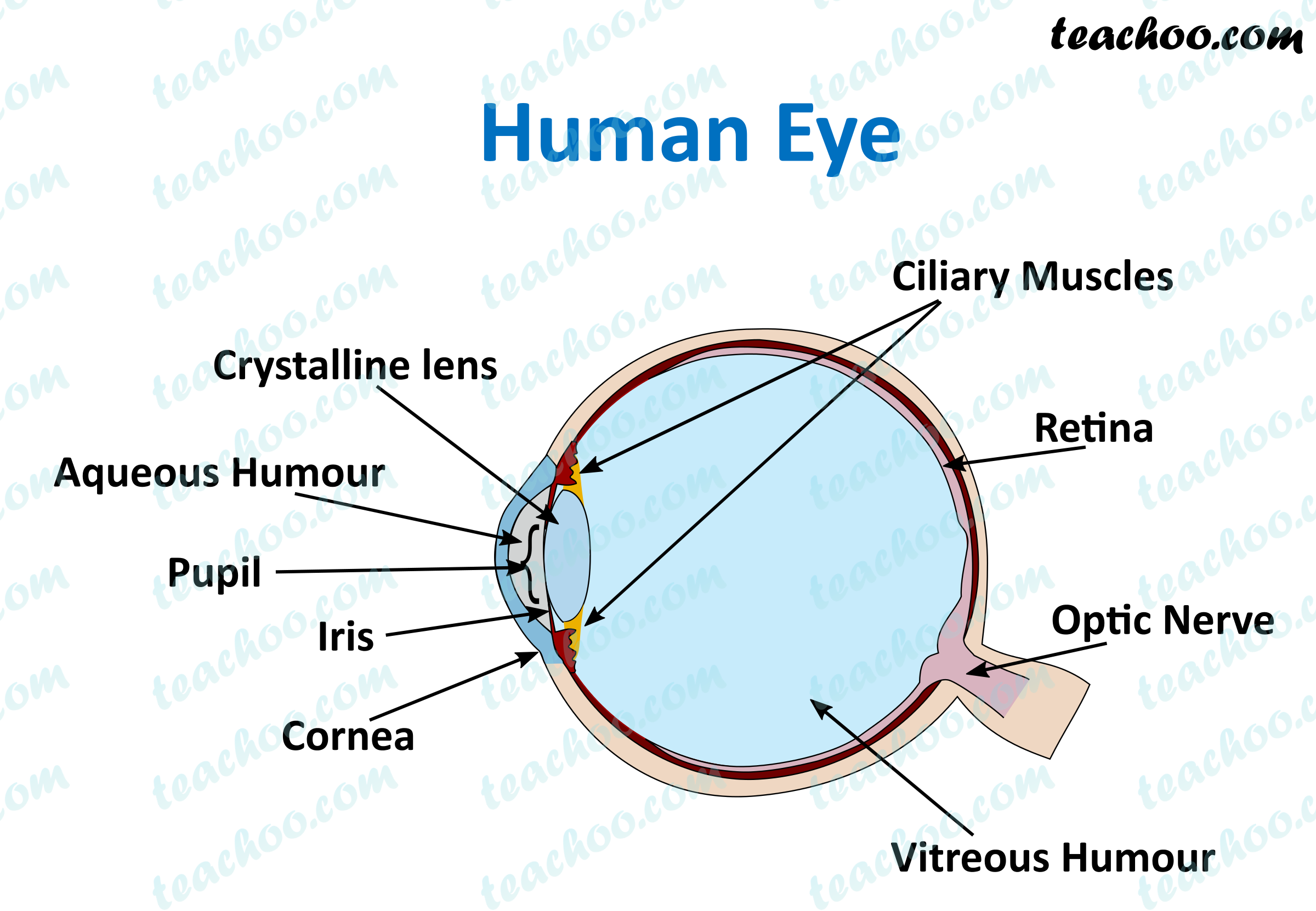
A problem with any of the three layers may cause dry eye.The accessory structures of the eye include the eyelids, eyelashes, eyebrows, the lacrimal apparatus, and extrinsic eye muscles. These create a film which covers the white of the eye and the cornea. Tears have three main components a watery component, an oily component and mucus. Vitreous humour – a clear gel that fills the inside of the eye and helps it to retain its shape. Sclera – also known as the white of your eye, this is the outer layer of the human eye. Retina – the light-sensitive tissue lining at the back of your eye that converts light into electrical impulses that are sent along the optical nerve to the brain. Pupil – this is the opening in the centre of the iris that lets in light. Optic nerve – a bundle of more than one million nerve fibres that carries visual messages from the retina to the brain. Macula – the sensitive area in the centre of the retina responsible for what you see ahead of you (central vision). Lens – the clear part of the eye behind the iris that helps to focus light, or an image, onto the retina. Iris – the coloured part of your eye that regulates the amount of light entering. Light rays pass through the pupil in the cornea.Īqueous humour – maintains the pressure in your eye and nourishes the cornea and the lens by supplying amino acids and glucose, as well as vitamin C.Ĭhoroid – a thin layer of blood vessels that nourish the retina and absorb scattered light.Ĭiliary muscles – a circular muscle that relaxes or tightens to enable the lens to change shape for focusing.Ĭornea – a clear covering on the front of your eye that focuses light entering the eye.įovea – a tiny pit in the macula that provides the sharp central vision that you need for activities, such as reading and driving. Find out about different parts of the eye.īasically, the role of the eye is to convert light into electrical signals called nerve impulses that the brain converts into images of our surroundings.


 0 kommentar(er)
